Three-point tail fiber
RCSB PDB
In this study, we have determined the structure of the alternative tail fiber subunit, gp52, and compared it with other tail fibers. The results revealed that Mu phage employs different structural motifs for two
Phage tail fibre assembly proteins employ a modular structure to drive
We demonstrate that Tfa forms a stable complex with the tail fibre, and present a 2.1 Å resolution X-ray crystal structure of this complex.
What Are Tail Fibers and Why Are They Important?
Tail fibers are structures on the phage that mediate their initial interaction with bacterial hosts, allowing them to recognize and attach to the bacterial surface.
Understanding Bacteriophage Tail Fiber
In this review, we comprehensively summarize how the tail fibers of the T4 phage recognize host surface receptors at single-molecule and atomic levels.
Structural Insights into the Chaperone-Assisted Assembly of a
In this study, we report the cryo-EM structure of the simplified tail fiber complexed with its chaperone from the myocyanophage Pam3, which provides insights into the assembly mechanism of the tail
Tail Fiber: Types, Functions, and Common Interfaces
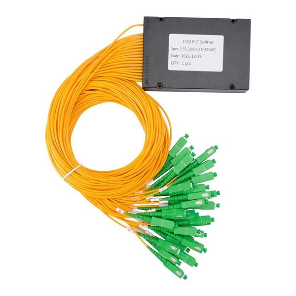
A tail fiber, also known as a fiber optic patch cord, consists of a connector on one end and a cut end of the fiber optic cable core on the other. These patch cords are primarily used to
Structural Insights into the Chaperone-Assisted
In this study, we report the cryo-EM structure of the simplified tail fiber complexed with its chaperone from the myocyanophage Pam3, which provides insights into
RBPseg: Toward a complete phage tail fiber structure atlas
Here, we introduce RBPseg, a method that combines monomeric ESMFold predictions with a structural-based domain identification approach, to divide tail fiber sequences into manageable
Architecture of the bacteriophage lambda tail: Structure
Its main role is to serve as an adaptor, connecting the 6-fold symmetric tail tube to the 3-fold symmetric central fiber protein (CFP). The CFP is attached to the BHP and is responsible for
Determination of the three-dimensional structure of bacteriophage Mu
The Mu phage can recognize lipopolysaccharide (LPS) on the host cell surface via its tail fibers. LPS is a component of the outer membrane of Gram-negative bacteria and consists of three
Asymmetric Structure of Podophage GP4 Reveals a Novel
In this study, we identified a new structure of the podophage with three types of tail fibers, and such phages with different types of fibers may have a broad host range and/or infect host cells
Frequently Asked Questions
- How to calculate the values of the distribution box
- Function of Outdoor Relay Protection Cabinets
- Sudan Flame-Retardant Fiber Optic Cable System Manufacturer for Smart Buildings
- Price of Canadian Cable Tray Screws
- How to connect two circuits to the distribution box
- Red and green labels on distribution boxes
- Singapore power distribution box manufacturers cost performance
- How to install an outdoor photovoltaic distribution box
- Customized optical modules available in stock
- Multimode fiber FC or SC
- Are high-voltage cable trays fireproof
- Costa Rican optical module manufacturer
- Spacing requirements between cable trays
- Remote Monitoring Solution for Romanian Hybrid Energy Systems
- QSFP28SFP Optical Module
- Custom Process for High-Temperature Resistant Fiber Optic Connectors for Security Applications
- Afghanistan Active Optical Module Smart RoHS
- Nordic warranty fiber optic Ethernet switch SFP
- How do high-speed optical modules transmit data
- Syrian imported optical cable splice box 2-core wholesale
- Price of QSFP Optical Line Terminal
- China Cable Tray Brackets
- Standard Height of Mobile Base Station Cable Tray